
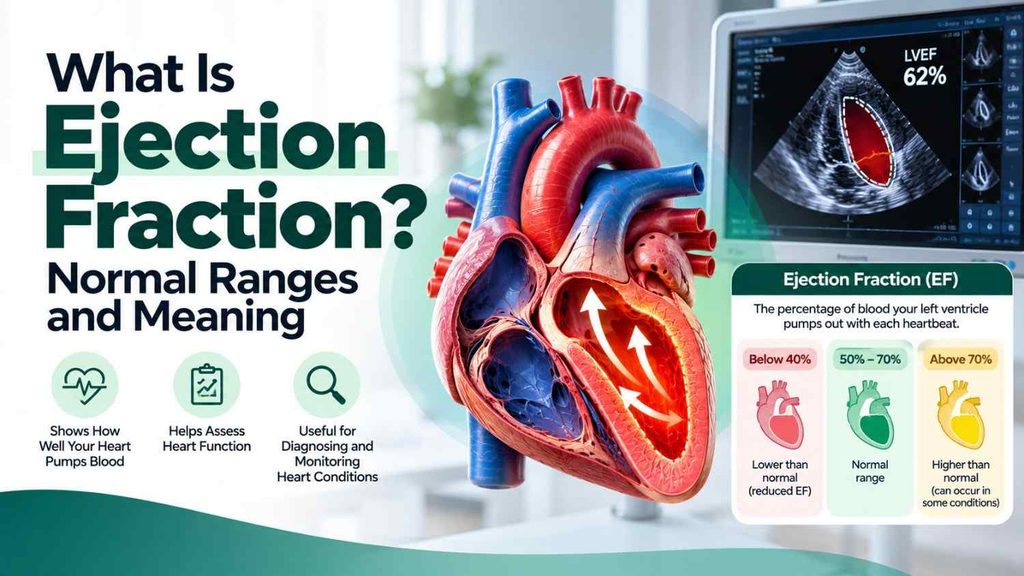
When you receive an echocardiogram report, you may notice a number followed by a percentage called the ejection fraction (EF). This measurement helps doctors understand how much blood the heart pumps out with each contraction. It is commonly used when evaluating heart function and certain types of heart failure.
But what is a normal ejection fraction? Does a low percentage always mean heart failure? And can someone have heart failure even when their EF is normal?
In general, a left ventricular ejection fraction of around 50% to 70% is considered within the normal range, although some medical references describe the typical range as approximately 55% to 70%. The interpretation depends on the measurement method, laboratory standards, and the person’s overall clinical condition.
This guide explains ejection fraction in simple terms, what different percentages may mean, how doctors measure it, and what questions to ask about your results. For broader information about cardiovascular conditions, visit our Heart Disease guide.
What Is Ejection Fraction?
Ejection fraction is the percentage of blood inside a heart ventricle that is pumped out with each heartbeat.
The heart has four chambers. The left ventricle is the main pumping chamber responsible for sending oxygen-rich blood to most of the body. Therefore, when people discuss ejection fraction, they are usually referring to left ventricular ejection fraction (LVEF).
For example, an EF of 60% means that the left ventricle pumps out approximately 60% of the blood it contains just before contraction. It does not mean that the heart pumps out 60% of all the blood in your body.
Some blood normally remains in the ventricle after each heartbeat. The heart fills again before the next contraction.
How Is Ejection Fraction Calculated?
Ejection fraction is calculated using the amount of blood in the ventricle before contraction and the amount pumped out during contraction.
The formula is:
Ejection fraction = (Blood pumped out ÷ Blood in the ventricle before contraction) × 100
For example, suppose the left ventricle contains 120 millilitres of blood before contraction and pumps out 72 millilitres.
EF = (72 ÷ 120) × 100 = 60%
This means the ejection fraction is 60%.
The actual assessment is performed using cardiac imaging or other specialised tests rather than by estimating these volumes yourself.
What Is the Normal Ejection Fraction Range?
A normal ejection fraction indicates that the ventricle is pumping out a typical proportion of the blood it contains. However, reference ranges can vary slightly between medical sources and testing methods.
The following categories are commonly used to explain left ventricular ejection fraction:
| Ejection fraction | General interpretation |
|---|---|
| Around 50%–70% | Generally considered within the normal range |
| 41%–49% | Mildly reduced or borderline, depending on the clinical context |
| 40% or lower | Reduced EF; may indicate significant impairment of pumping function |
| Above approximately 70%–75% | May be normal in some circumstances or require further interpretation if unusually high |
These are general guideposts, not a diagnosis. Your clinician will interpret your EF alongside your symptoms, imaging findings, medical history, and the reference range used by the testing facility.
An EF of 45%, for example, should not automatically be interpreted as heart failure without considering the rest of the clinical assessment. Similarly, an EF within the normal range does not exclude every form of heart failure.
What Does a Low Ejection Fraction Mean?
A low ejection fraction means that the left ventricle is pumping out a smaller proportion of its blood than expected. It may indicate that the heart muscle is not contracting effectively, although the cause and significance vary.
Doctors may describe an EF of 41%–49% as mildly reduced and an EF of 40% or less as reduced. These ranges are commonly used in the classification of heart failure, but a diagnosis requires more than the EF number alone.
Possible Causes of Low Ejection Fraction
Several conditions may contribute to a reduced EF, including:
- Coronary artery disease: Narrowed or blocked coronary arteries can reduce the blood supply to the heart muscle.
- Previous heart attack: Damage to heart muscle may weaken its ability to contract.
- Cardiomyopathy: Diseases of the heart muscle can affect its strength and function.
- Heart valve disease: Certain valve problems can place extra strain on the heart.
- Inflammation of the heart muscle: Conditions such as myocarditis can impair pumping function.
- Persistent abnormal heart rhythms: Some prolonged rhythm disturbances can weaken the heart over time.
- Other medical conditions: Certain systemic illnesses, treatments, or inherited conditions may affect heart function.
The cause cannot be determined from the EF percentage alone. Additional assessment may be needed to identify the underlying condition.
Symptoms That May Occur With Low EF
Some people with reduced EF experience symptoms, while others have few or no noticeable symptoms, particularly in the early stages.
Possible symptoms include:
- Shortness of breath during activity or while lying down
- Unusual tiredness or reduced exercise tolerance
- Swelling in the feet, ankles, or legs
- Rapid or irregular heartbeat
- Persistent coughing or difficulty breathing when lying flat
- Unexpected weight gain caused by fluid retention
These symptoms can have causes other than heart failure. Seek medical assessment if they are persistent, new, or worsening.
Severe or sudden shortness of breath, chest pain, fainting, or signs of a medical emergency require urgent care.
What Does a Normal Ejection Fraction Mean?
A normal EF generally indicates that the left ventricle pumps out a typical proportion of its blood with each contraction. It is reassuring in the context of many assessments, but it does not tell the whole story about heart health.
A person can have a normal EF and still have a cardiovascular condition that needs treatment.
For example, in heart failure with preserved ejection fraction (HFpEF), the heart muscle may become stiff and fail to relax or fill properly. The ventricle can pump out a normal proportion of the blood it contains, yet the amount of blood available to pump may be insufficient to meet the body’s needs during activity or at rest.
A normal EF also does not rule out coronary artery disease, heart valve problems, abnormal heart rhythms, or other conditions.
This is why doctors interpret EF alongside symptoms, physical examination findings, and other tests rather than treating it as a standalone measure of overall heart health.
What Does a High Ejection Fraction Mean?
A higher-than-expected EF does not automatically mean that the heart is healthier or stronger.
An EF above the usual reference range may occur for different reasons, including differences in how the heart fills and contracts or certain heart muscle conditions. In some situations, a high EF may be associated with a small, stiff ventricle that pumps out a high percentage of a relatively small amount of blood.
The significance depends on the person’s symptoms, heart structure, and other findings.
If your report shows an unusually high EF, ask your clinician whether it is normal for your circumstances or whether further evaluation is appropriate. Do not assume that a higher percentage is always better.
How Is Ejection Fraction Measured?
Doctors can estimate or measure EF using several tests. The choice depends on the clinical question, the quality of the images, and the person’s medical circumstances.
1. Echocardiogram
An echocardiogram is one of the most commonly used tests for measuring ejection fraction. It uses ultrasound waves to create images of the heart, allowing clinicians to assess its chambers, movement, valves, and pumping function.
The test is generally non-invasive and does not use ionising radiation. The report may include EF along with other measurements of heart structure and function.
Learn more in our guide to the echocardiogram. Ask your clinician which specific imaging test is appropriate if you need a more detailed explanation of the procedure.
2. Cardiac MRI
Cardiac magnetic resonance imaging uses a magnetic field and radio waves to produce detailed images of the heart. It can measure ventricular volumes and EF and may help investigate heart muscle disease when additional detail is needed.
Not everyone needs a cardiac MRI. It may be recommended when an echocardiogram does not provide enough information or when doctors need to assess a particular heart condition.
3. Nuclear Cardiac Imaging
Certain nuclear imaging tests can estimate ventricular function and ejection fraction. One example is a multigated acquisition scan, often called a MUGA scan.
These tests may be used in selected circumstances, depending on the clinical question and the availability of alternatives.
4. Cardiac Catheterisation or Other Imaging
Some procedures and imaging methods can also assess ventricular function. The appropriate test depends on why EF needs to be measured and what other information the clinician needs.
An ECG, by contrast, records the heart’s electrical activity. It can help identify rhythm problems and other signs of heart disease, but it does not directly measure EF.
How Accurate Is an Ejection Fraction Result?
EF is an estimate, and results can vary depending on the imaging method, image quality, the person performing the measurements, and changes in the person’s condition.
Different tests may produce slightly different EF values. Even repeated assessments using the same method may vary.
When comparing two reports, consider:
- Whether the same imaging method was used
- The dates of the tests
- Whether image quality was adequate
- Whether there have been changes in symptoms or treatment
- The other findings described in each report
A small change in EF does not always mean that heart function has meaningfully improved or worsened. Your clinician can determine whether a difference is clinically important.
Can Ejection Fraction Improve?
Yes. In some people, a reduced EF can improve when the underlying cause is treated and appropriate care is provided. The degree of improvement depends on the cause, how much heart muscle damage has occurred, the person’s overall health, and the response to treatment.
Management may include:
- Treating the underlying cause: For example, addressing coronary artery disease or certain heart valve problems when indicated.
- Prescribed medication: Specific medicines can improve outcomes in appropriate forms of heart failure and may help improve EF.
- Managing blood pressure and diabetes: Controlling these conditions can reduce strain on the heart.
- Following an appropriate activity plan: Physical activity and cardiac rehabilitation may be recommended according to the person’s condition.
- Making sustainable lifestyle changes: Avoiding tobacco, following an appropriate diet, and limiting alcohol when advised can support cardiovascular health.
- Regular follow-up: Repeat imaging may be recommended to monitor changes in heart function.
Do not start, stop, or adjust heart medicines based on an EF number alone. Some people need long-term treatment even after their EF improves, so decisions should be made with their treating clinician.
When Should You See a Doctor About Your EF Result?
Discuss your report with a healthcare professional if:
- Your EF is below the reference range stated in the report.
- You have new or worsening breathlessness, fatigue, or swelling.
- You have been diagnosed with heart failure and need help understanding your results.
- Your EF has changed significantly between tests.
- You have a history of heart attack, cardiomyopathy, or significant valve disease.
Your clinician may review your symptoms, medical history, physical examination, and other test results to determine what the number means for you.
If you develop severe chest pain, sudden severe breathlessness, collapse, or other signs of an emergency, seek emergency medical care immediately rather than waiting for a routine appointment.
Frequently Asked Questions
1. What is a normal ejection fraction for adults?
A left ventricular ejection fraction of approximately 50%–70% is commonly considered normal, although some references use a range of around 55%–70%. The correct interpretation depends on the measurement method and clinical context.
2. Is an ejection fraction of 55% good?
An EF of 55% is generally within the normal range in many clinical references. However, it should be interpreted alongside symptoms and other heart findings. A normal EF does not exclude every form of heart disease or heart failure.
3. Is an ejection fraction of 40% dangerous?
An EF of 40% is considered reduced and may indicate impaired pumping function. Its significance depends on the cause, symptoms, and other findings. Arrange a clinical review to understand your result and treatment needs; seek urgent care for severe or sudden symptoms.
4. Can you have heart failure with a normal ejection fraction?
Yes. Heart failure with preserved ejection fraction occurs when the heart has difficulty relaxing or filling adequately, even though the percentage of blood pumped out remains in the preserved range. Doctors use symptoms, examination findings, and other tests to establish the diagnosis.
5. Does a higher ejection fraction mean a healthier heart?
Not necessarily. A high EF is not always better, and an unusually high value may need interpretation alongside heart structure and filling. Overall heart health cannot be judged from EF alone.
6. Which test measures ejection fraction most commonly?
An echocardiogram is one of the most commonly used tests. Cardiac MRI, nuclear imaging, and certain other procedures can also measure or estimate EF when appropriate.
7. Can a low ejection fraction return to normal?
It can improve in some people, depending on the underlying cause and response to treatment. Medication, treatment of the underlying condition, lifestyle changes, and follow-up care may help. Improvement is not guaranteed, and treatment decisions should be individualised.
8. How often should ejection fraction be checked?
There is no single schedule for everyone. Repeat testing depends on the diagnosis, symptoms, treatment changes, and the clinician’s assessment. Ask your healthcare professional when another echocardiogram or other imaging test is appropriate.
Conclusion
Ejection fraction is an important measurement that helps doctors understand how much blood the left ventricle pumps out with each heartbeat. A value around 50%–70% is commonly considered within the normal range, while lower results may indicate reduced pumping function.
However, EF is only one part of a complete heart assessment. Some people have heart failure despite a preserved EF, and an unusually high EF does not automatically mean better heart health.
If your report shows an abnormal result, discuss it with a qualified healthcare professional rather than interpreting the percentage in isolation. Understanding the number alongside your symptoms, medical history, and other test findings can help guide appropriate care.
For more information about cardiovascular conditions, visit the Heart Disease guide at Ghaziabad Clinic.