
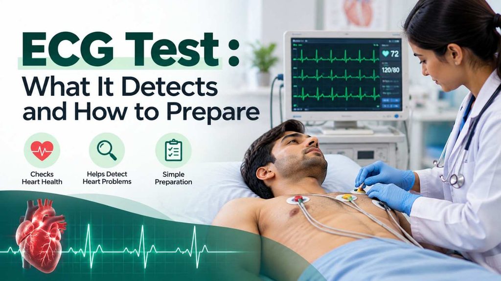
An ECG test (electrocardiogram) records the electrical signals that control your heartbeat. Doctors use this test to evaluate heart rhythm, identify certain electrical abnormalities, and look for signs that may point to an underlying heart condition.
An ECG is commonly recommended when someone experiences a racing heartbeat, chest discomfort, dizziness, or unexplained changes in exercise tolerance. It may also be used to monitor an existing heart condition or check how certain treatments affect the heart.
The procedure is usually quick, painless, and non-invasive. However, understanding what an ECG can and cannot reveal is important because a normal tracing does not rule out every heart problem.
In this guide, you will learn what an ECG test detects, how to prepare, what happens during the procedure, how results are interpreted, and when another heart test may be necessary.
What Is an ECG Test?
An electrocardiogram, also called an ECG or EKG, is a diagnostic test that records the electrical activity generated by your heart. The terms ECG and EKG refer to the same test.
Each heartbeat begins with an electrical impulse that travels through the heart’s specialised conduction system. This signal coordinates the contraction of the heart chambers, helping blood move through the lungs and the rest of the body.
During an ECG test, small adhesive sensors called electrodes are placed on the skin, usually across the chest and on the arms and legs. Wires connect these electrodes to a recording device, which converts the electrical signals into a tracing displayed on a screen or printed on paper.
A healthcare professional evaluates the tracing to assess the heart’s rate, rhythm, electrical conduction, and other patterns that may require further investigation.
Unlike an ultrasound scan, an ECG does not create pictures of the heart’s internal structures. It records electrical activity instead.
What Does an ECG Test Detect?
The main purpose of an ECG is to assess the heart’s electrical activity. Its findings can help doctors identify certain abnormalities and decide whether additional investigations are necessary.
1. Abnormal Heart Rhythms
An ECG can identify many heart rhythm disorders, also known as arrhythmias. These occur when the heart beats too quickly, too slowly, or irregularly.
Depending on the rhythm and whether it is present during recording, an ECG may reveal atrial fibrillation, certain forms of tachycardia, bradycardia, or other electrical abnormalities.
Some rhythm disturbances occur intermittently. If they are absent during a brief ECG recording, the test may not capture them.
Learn more about heart arrhythmia and how abnormal electrical signals affect the heartbeat.
2. Possible Signs of a Heart Attack
An ECG can reveal electrical changes associated with an ongoing or previous heart attack. These findings can help clinicians assess a person with suspected acute coronary problems.
However, a normal ECG does not completely exclude a heart attack. Symptoms, examination findings, blood tests such as cardiac troponin, and sometimes repeat ECGs or imaging are needed to reach a diagnosis.
If you have new chest pressure, severe breathlessness, or other possible heart attack symptoms, seek emergency medical care rather than waiting for a routine ECG appointment.
3. Electrical Conduction Problems
The heart uses an organised electrical pathway to coordinate each beat. An ECG can reveal delays or blocks in the transmission of electrical impulses.
For example, it may identify certain atrioventricular blocks or bundle branch blocks. The significance depends on the specific pattern, symptoms, medical history, and other test results.
4. Clues to Heart Muscle or Chamber Abnormalities
Some ECG patterns can suggest that the heart muscle has thickened, that a chamber may be enlarged, or that the heart’s electrical pattern has changed because of an underlying condition.
An ECG cannot reliably measure all structural changes. If a doctor needs to examine chamber size, valve function, or pumping performance, an echocardiogram or another imaging test may be more appropriate.
5. Changes That May Be Associated With Reduced Blood Supply
Certain ECG changes may suggest that the heart muscle is not receiving enough blood or oxygen. These findings can occur in coronary artery disease or during an acute coronary event.
An ECG alone cannot determine whether a coronary artery is narrowed or blocked in every case. Further assessment may involve blood tests, stress testing, coronary CT angiography, or other investigations based on the clinical situation.
Read our guide to coronary artery disease for more information about narrowed arteries supplying the heart.
6. Effects of Certain Medicines or Electrolyte Imbalances
Some medicines can affect the heart’s electrical conduction or the time it takes for electrical signals to recover between beats. Changes in blood electrolytes, particularly potassium or calcium, can also alter the ECG pattern.
When these changes are suspected, doctors may review your medication list and order blood tests to investigate the cause.
7. Monitoring an Existing Heart Condition
An ECG may be repeated to monitor a known rhythm disorder, assess the effects of selected medicines, or check electrical activity after a cardiac procedure or pacemaker placement.
Comparing a current tracing with earlier ECGs can help clinicians identify changes over time.
When Should You Get an ECG Test?
A doctor may recommend an ECG when symptoms or medical history suggest a possible heart problem.
Common reasons include:
- Chest discomfort or pain that needs medical evaluation.
- A racing, pounding, or irregular heartbeat.
- Dizziness, fainting, or unexplained episodes of light-headedness.
- Shortness of breath or reduced ability to exercise.
- Suspected heart rhythm or conduction abnormalities.
- Follow-up of a previously diagnosed heart condition.
- Monitoring when a medicine may affect heart rhythm.
- Assessment before certain operations when clinically indicated.
An ECG may also be part of an urgent assessment in a hospital or emergency department.
Not everyone needs an ECG as a routine screening test. Whether it is useful for someone without symptoms depends on individual risk factors, medical history, and the reason for considering the test.
What Are the Different Types of ECG Tests?
The type of ECG recommended depends on when symptoms occur and what the doctor needs to investigate.
Resting ECG Test
A resting ECG is the standard form of electrocardiogram. You lie comfortably while the machine records your heart’s electrical activity for a short period.
It is often used to investigate symptoms, assess a suspected rhythm problem, or establish a baseline tracing.
Exercise ECG or Stress Test
An exercise ECG records the heart’s electrical activity while you walk on a treadmill or pedal a stationary bicycle. Your heart rate and blood pressure may also be monitored.
The test helps doctors assess how your heart responds to physical exertion. It may be used for selected people with suspected coronary artery disease or exercise-related symptoms.
If you cannot exercise adequately, a clinician may consider another type of investigation, including a pharmacological stress test. Not everyone is suitable for exercise ECG testing.
Holter Monitor
A Holter monitor is a portable device that records the heart’s electrical activity continuously, often for 24 to 48 hours, although monitoring duration varies by device and clinical need.
It may be recommended when symptoms such as palpitations occur repeatedly but are not present during a standard ECG.
You can usually continue many ordinary daily activities while wearing the device, following the instructions provided by the monitoring team.
Event Monitor or Longer-Term ECG Monitoring
An event monitor or another ambulatory ECG device records heart rhythm over a longer period. Some devices record continuously, while others capture or transmit information when an event occurs or an abnormal rhythm is detected.
These monitors may be useful when symptoms happen infrequently and a short recording is unlikely to capture them.
Your doctor will select the monitoring method based on how often symptoms occur and what needs to be investigated.
How to Prepare for an ECG Test
Preparing for a standard resting ECG is usually straightforward. Most people do not need to fast or follow a special diet.
The following steps can help the test proceed smoothly.
1. Wear Comfortable Clothing
Choose clothing that allows easy access to your chest, wrists, and ankles. You may be asked to remove or adjust clothing covering the areas where electrodes will be placed. A gown or covering may be provided for privacy.
2. Keep Your Skin Clean
Avoid applying body lotion, oil, or powder to the areas where the electrodes will be attached. These products may make it harder for the sensors to adhere properly.
If you have significant chest hair, the technician may need to trim a small area to ensure good contact.
3. Tell the Healthcare Team About Your Medicines
Provide an accurate list of prescription medicines, over-the-counter products, and supplements you take.
Some medicines affect heart rate or electrical conduction, which may influence the findings. Do not stop or change any medicine unless your clinician specifically instructs you to do so.
4. Ask Whether Special Instructions Apply
A standard resting ECG usually requires no fasting. However, an exercise ECG or another specialised test may have different preparation requirements.
For a stress test, you may receive instructions about meals, caffeine, exercise, and medicines. Follow the instructions from the facility performing the test rather than applying resting-ECG preparation rules to every type.
5. Inform the Team About Relevant Medical Details
Tell the technician or clinician if you have a pacemaker, an implanted cardiac device, a skin condition affecting electrode placement, or a previous ECG that may be useful for comparison.
Mention your symptoms and when they tend to occur. This information may help your doctor decide whether a standard ECG is sufficient or longer monitoring is needed.
What Happens During an ECG Test?
A resting ECG is generally completed within a few minutes. The overall appointment may take longer depending on preparation and the facility’s process.
Here is what you can usually expect.
Step 1: Getting ready
You will be asked to lie on an examination bed. The technician may clean selected areas of your skin to help the electrodes adhere securely.
Step 2: Attaching the electrodes
Small adhesive sensors are placed on the chest and limbs. A standard 12-lead ECG uses electrodes positioned in specific locations to record the heart’s electrical activity from multiple perspectives.
The electrodes are connected to the ECG machine by wires or leads.
Step 3: Recording the tracing
You will be asked to lie still, relax, and breathe normally. Movement, muscle tension, or shivering can interfere with the recording.
The machine records the electrical signals and displays them as a series of waves.
Step 4: Removing the sensors
Once the recording is complete, the electrodes are removed. You can usually return to your normal activities immediately after a resting ECG unless your clinician gives different instructions.
The recording is then reviewed by an appropriately qualified healthcare professional.
How Long Does an ECG Test Take?
A standard resting ECG recording usually takes only a few minutes. Allow additional time for registration, electrode placement, and any discussion with the healthcare team.
An exercise ECG takes longer because your heart activity is recorded during exercise and recovery. Holter monitoring and other ambulatory ECG tests last much longer, ranging from hours to days or sometimes weeks, depending on the device and the reason for testing.
The duration of the test should not be confused with the time required to receive or discuss the final report. That varies by facility and clinical urgency.
Is an ECG Test Painful or Risky?
A standard ECG is non-invasive and generally painless. It does not send electricity through your body; it only records the electrical signals produced by your heart.
You may feel the adhesive electrodes being attached or removed. Occasionally, they cause mild skin irritation, particularly in people with sensitive skin.
A resting ECG has very little risk. Exercise ECG testing involves physical exertion, so it requires appropriate patient selection and supervision. The clinical team will explain any relevant precautions before a stress test.
How to Understand ECG Test Results
An ECG tracing contains electrical waves and intervals that healthcare professionals assess in context. The report should be interpreted alongside your symptoms, medical history, examination, and any other investigations.
What Does a Normal ECG Mean?
A normal ECG generally indicates that the recorded heart rate, rhythm, and electrical patterns do not show significant abnormalities detectable by that tracing.
However, a normal result does not guarantee that the heart is completely healthy. Some conditions, including intermittent rhythm disorders and certain forms of coronary artery disease, may not produce noticeable changes during a short recording.
If your symptoms continue despite a normal ECG, your doctor may recommend additional testing.
What Does an Abnormal ECG Mean?
An abnormal ECG means that one or more features of the electrical tracing differ from the expected pattern. This does not automatically mean you have a serious heart condition.
Possible explanations include:
- A fast, slow, or irregular rhythm.
- Delayed electrical conduction.
- Changes associated with a previous or current heart problem.
- Effects of certain medicines or electrolyte abnormalities.
- Variations related to body position, electrode placement, or recording artefacts.
The clinical importance depends on the specific abnormality. Some findings require urgent attention, while others may be longstanding or need only further evaluation.
What Are P Waves, QRS Complexes, and T Waves?
You may notice these terms on an ECG report. They describe components of the electrical tracing.
- P wave: Represents electrical activation of the upper heart chambers, called the atria.
- QRS complex: Represents electrical activation of the lower chambers, called the ventricles.
- T wave: Represents the electrical recovery of the ventricles after activation.
Clinicians also assess intervals, the relationship between waves, rhythm patterns, and other features. These measurements are not intended to be interpreted in isolation by patients.
If you receive an ECG report, ask your healthcare professional to explain the findings in relation to your symptoms and medical history.
Can an ECG Detect All Heart Problems?
No. An ECG is a valuable diagnostic tool, but it does not detect every heart condition.
A resting ECG provides a short recording of electrical activity. It may not capture a rhythm problem that occurs only occasionally. It also does not directly show the heart’s valves, blood flow through the coronary arteries, or the full structure of the heart muscle.
Depending on the suspected condition, your doctor may recommend other tests.
- Echocardiogram: Uses ultrasound to assess the heart’s structure, valves, and pumping function.
- Blood tests: May identify cardiac injury or other medical abnormalities. Cardiac troponin is particularly important when a heart attack is suspected.
- Holter or event monitoring: Records electrical activity over a longer period to investigate intermittent symptoms.
- Exercise or other stress testing: Assesses how the heart responds to exertion or medication-induced stress in selected patients.
- Cardiac imaging: CT, MRI, or other imaging methods may be used to investigate specific structural or coronary problems.
The most appropriate test depends on the clinical question, not simply on whether an ECG is normal or abnormal.
Frequently Asked Questions About ECG Tests
1. Is an ECG test the same as an EKG?
Yes. ECG and EKG are two abbreviations for electrocardiogram. Both refer to a test that records the electrical activity of the heart.
2. Do I need to fast before an ECG test?
Usually, no. A standard resting ECG generally does not require fasting. Exercise ECG and other specialised tests may have different instructions, so check with the facility conducting your test.
3. Can I eat or drink before an ECG?
For a standard resting ECG, you can generally eat and drink normally. If you are scheduled for an exercise or specialised cardiac test, follow the specific preparation instructions provided by your healthcare team.
4. Can an ECG detect a heart attack?
An ECG can reveal changes that suggest a heart attack, but it cannot rule out every heart attack on its own. Doctors may also use symptoms, repeat ECGs, cardiac troponin blood tests, and other investigations.
5. Can an ECG detect blocked arteries?
An ECG may show changes associated with reduced blood supply to the heart, but it cannot reliably identify or exclude all coronary artery blockages. Additional tests may be necessary when coronary artery disease is suspected.
6. Is an ECG safe during pregnancy?
A standard ECG does not use ionising radiation and is generally considered safe during pregnancy when medically indicated. Inform your healthcare professional that you are pregnant so your care can be appropriately tailored.
7. Why might I need another ECG after a normal result?
A short ECG may not capture symptoms that occur intermittently. If palpitations, dizziness, chest discomfort, or other concerns continue, your doctor may recommend a repeat ECG, longer-term monitoring, or another investigation.
8. Who interprets an ECG report?
An appropriately qualified healthcare professional interprets the tracing in the context of the person’s clinical history and symptoms. Automated machine interpretations can be helpful, but they should not replace appropriate clinical review.
9. When should I seek urgent medical attention?
Seek emergency medical care if you have new or severe chest pain or pressure, severe breathlessness, collapse, or other symptoms that could indicate a heart attack or another serious emergency. Do not wait for a scheduled ECG or a report before seeking help.
Conclusion
An ECG test provides important information about the heart’s electrical activity and can help doctors investigate abnormal rhythms, conduction problems, and certain changes associated with heart disease.
Preparing for a standard ECG is usually simple, and the procedure is generally quick and painless. However, the results must be interpreted alongside your symptoms and medical history because a normal tracing does not exclude every heart condition.
If your doctor recommends an ECG, ask what they are investigating and whether any additional tests may be needed. If symptoms persist or worsen, seek medical advice even if an earlier ECG was reported as normal.